About Diagnostic Testing
Optical Coherence Tomography
Optical Coherence Tomography, or OCT, is a non-invasive, fast-imaging, scanning technology that utilizes coherent light beams to document retinal structures or pathology. As it scans the macula, a detailed and magnified cross sectional image is produced which is used to document retinal structures and pathology. The retinal thickness is also measured which can aid in the diagnosis and monitoring of many retinal diseases. OCT usually takes only a few minutes.

Fundus Photography
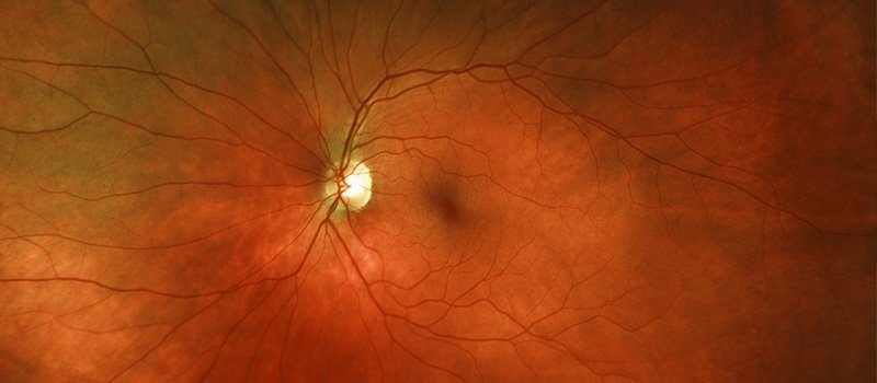
Digital color photographs are taken of the retina using specialized fundus cameras. The term fundus describes the back of the eye including the retina and the optic nerve. This non-invasive procedure allows the retinal specialist to document retinal structures or pathology which can be used for future comparison. These photographs are commonly combined with fluorescein angiography to further evaluate retinal conditions. Fundus photography usually takes about five to ten minutes.
Fluorescein Angiography
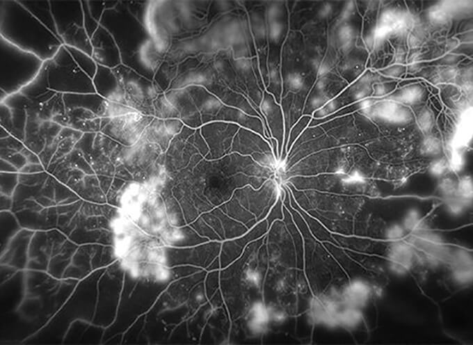
Fluorescein angiography is an imaging technique that highlights the circulation of the retina using a series of rapid-sequence photographs or video imaging. Fluorescein sodium, a diagnostic dye, is injected through a small butterfly needle into a vein in the arm. Within seconds, a series of photographs or video is taken to document the circulation of the dye through the arteries and veins in the retina. Leakage, blockage and other vascular abnormalities may be disclosed which can aid in the diagnosis and evaluation of many retinal conditions. Although complications and side effects are rare with fluorescein sodium they include inflammation, pain or bruising at the injection site and more rarely, itching, hives, breathing difficulties or even more severe allergic reactions. The fluorescein sodium dye may give a yellowish tint to the patient’s skin and it usually turns urine a bright yellow or orange color for 24 to 36 hours. If undergoing fluorescein angiography patients are urged to alert their physician and photographer of any know allergies to iodine, shellfish or contrast dyes. Fluorescein angiography usually takes about fifteen to twenty minutes.
Indocyanine Green Angiography
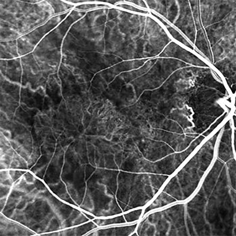
Indocyanine Green Angiography, or ICG, is much like fluorescein angiography with the addition of a green cyanine dye. Both of the contrast dyes, fluorescein sodium and indocyanine green, are injected intravenously followed by rapid sequence photographs or video. ICG angiography can help identify conditions and abnormalities within the vascular layer located beneath the retina called the choroid. Possible complications and side effects with ICG angiography are similar to those found with fluorescein angiography. If undergoing ICG angiography patients are urged to alert their physician and photographer of any known allergies to iodine, shellfish or contrast dyes. ICG angiography usually takes about fifteen to twenty minutes
Autofluorescence Photography
Autofluorescence photography is non-invasive, digital imaging which uses specialized cameras to identify retinal pathology. Certain retinal abnormalities appear to be fluorescent when illuminated with blue light then photographed using filters. Autofluorescence imaging is used to evaluate and document damaged areas of the retina so they can be monitored over time. Autofluorescence photography usually takes about five to ten minutes.
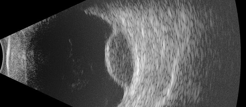
Standardized A&B-scan Echography
A&B-Scan ultrasound imaging (echography) is a non-invasive technology which uses high frequency sound waves to scan internal eye structures. During the ultrasound exam, sound waves are used to produce echoes which are reflected from the back of the eye. These echoes are used to create a two-dimensional image which displays both normal and abnormal ocular structures. Ultrasound imaging is useful when the back of the eye cannot be adequately viewed by a physician due to conditions such as vitreous hemorrhage, dense cataracts, or any pathology which blocks the view of the internal eye structures. Ultrasound testing may take about ten to fifteen minutes per eye.
Perimetry (Visual Field Testing)
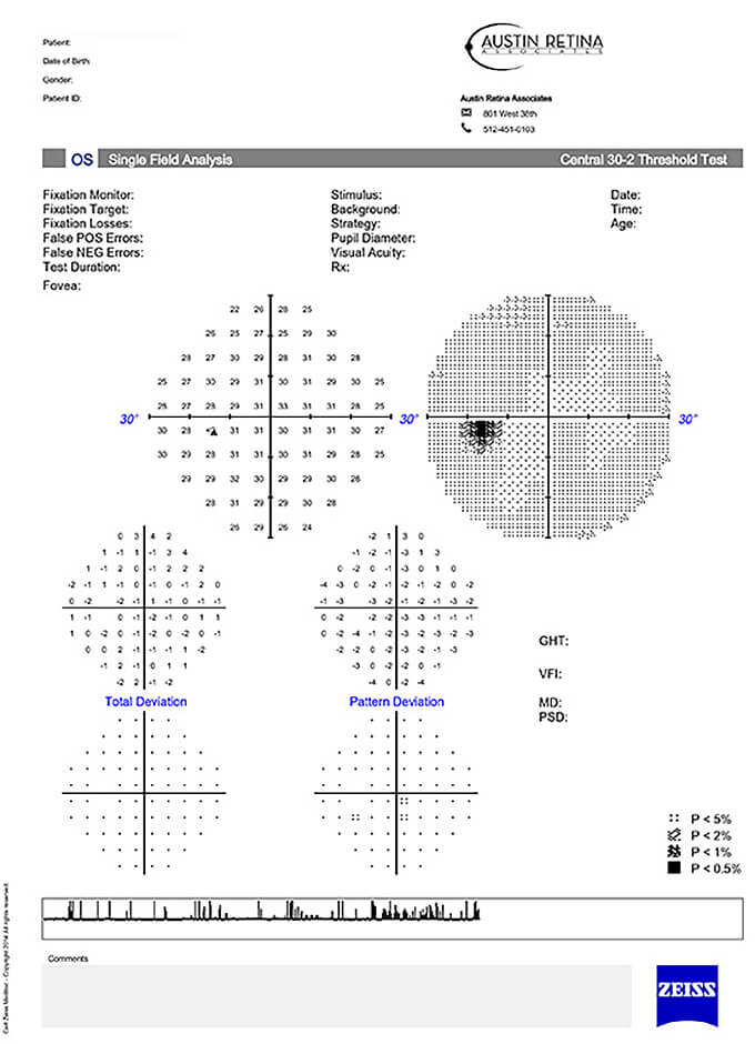
Perimetry, or visual field testing, is a non-invasive test of the central and peripheral fields of vision. During visual field testing an automated or manual perimeter displays a series of small lights randomly in front of each eye. The patient responds by pressing a button each time a test light is seen. The visual field test maps the extent of a patient’s central and peripheral vision as well as documents abnormalities such as blind spots or missing areas of vision which may indicate the presence of a retinal, optic nerve or visual pathway condition. Visual field testing often takes only five minutes per eye but may take as long as thirty minutes per eye.
