About Choroidal Melanoma
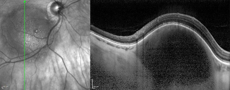
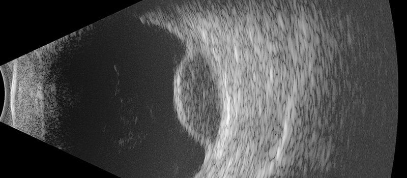
Uveal melanomas are tumors of pigmented cells and often arise from choroidal nevi (also known as “moles”) inside the eye. Depending upon the location and size of the melanoma, patients may have very few symptoms, or they may notice peripheral visual field loss or blurring of the central vision.
Diagnosis of uveal melanoma is made through a combination of dilated examination, ophthalmic photos, ultrasonography, and other forms of imaging such as optical coherence tomography and fluorescein angiography. Unlike other forms of cancer, the diagnosis is usually made based upon the exam and imaging and without a biopsy. In cases where the tumor is atypical, a biopsy may be performed to help confirm the diagnosis, and genetic testing of the tumor can also be performed at this time. Part of the initial diagnostic workup includes imaging of the body with PET/CT, CT, or MRI to see if the tumor may have spread to other parts of the body.

The primary goal of treatment of uveal melanoma is to prolong life by preventing metastatic spread to other parts of the body. The main treatments for uveal melanoma are radiation treatment with plaque brachytherapy for smaller tumors and enucleation (removal of the eye) for larger tumors. For certain tumors, proton beam therapy (another form of radiation) or laser treatment (with transpupillary thermotherapy) may be recommended. The benefit of plaque brachytherapy is that the eye can be preserved, and vision may also be preserved depending upon the location and size of the tumor. The risks of plaque radiotherapy include vision loss (particularly if the tumor is close to the center of the retina or the optic nerve), the risk of spread to other parts of the body, the risk of need for additional treatment if there is tumor recurrence within the eye (which can include laser treatment or enucleation), high eye pressure, cataract, and double vision after the surgery. It is important to continue screening for spread of the melanoma to other parts of the body even after the eye treatment has been completed.

Plaque brachytherapy requires two surgeries, one to place the plaque against the eye over the area of the tumor, and a second surgery to remove the plaque approximately one week later. Patients may experience eye redness, swelling, and a sensation of fullness while the plaque is in place. After treatment, periodic visits are required to monitor the response of the tumor within the eye, as well as continued screening for spread to other parts of the body.
For additional information, please visit the ASRS page on choroidal melanoma.

